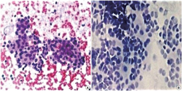
Fig. 1.

(A) Fine needle aspiration cytology of neuroblastoma showing sheets and clusters of round, monomorphic tumor cells on a hemorrhagic background. (B) Non-aspiration cytology of neuroblastoma showing clusters and dispersed small, round cells with a high nucleocytoplasmic ratio and scant cytoplasm.
