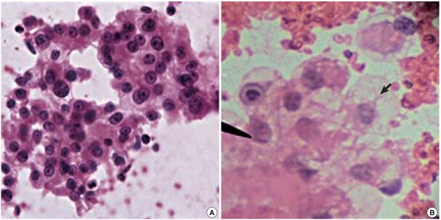
Fig. 2.

(A) Fine needle cytology with non-aspiration smear of renal cell carcinoma showing sheets and clusters of cells with abundant, delicate, wispy, finely vacuolated cytoplasm and enlarged nuclei, fine chromatin, prominent nucleoli and thick irregular nuclear border on a relatively clean background. (B) Fine needle cytology with aspiration smear of renal cell carcinoma showing a sheet of cells with abundant vacuolated cytoplasm (arrow) and enlarged nuclei on a haemorrhagic background.
