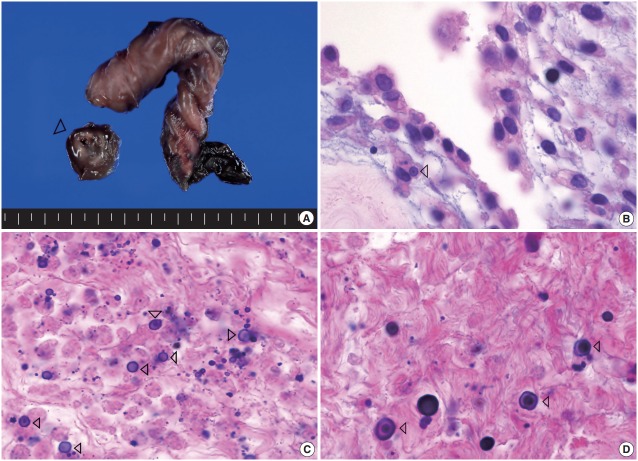
Fig. 1.
Representative macroscopic and microscopic images of the lesion. (A) The middle portion of umbilical cord is focally enlarged, inside which an ill-defined white to brown horseshoe-shaped area (arrowhead) surrounding the umbilical vessels is noted. (B) The concentrically laminated, round-ovoid, basophilic calcified bodies, referred to as Michaelis-Gutmann bodies, are seen within macrophages (arrowhead). (C) This lesion is composed of multiple, small, round to oval, calcified bodies (calcospherites), Michaelis-Gutmann bodies (arrowheads), and an inflammatory infiltrate containing mainly necrotic macrophages. (D) The Michaelis-Gutmann bodies (arrowheads) are also observed in the noncalcified area.

