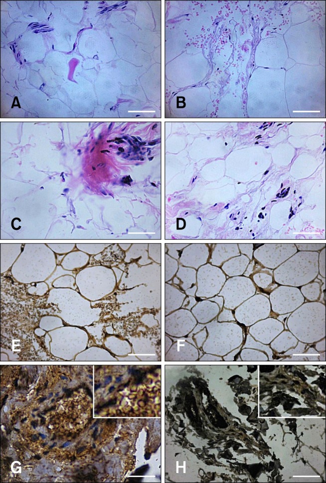
Fig. 1. Histology and immunohistochemistry examination of adipose tissue collected from equine metabolic syndrome (EMS; group A; panels A, C, E, and G) and healthy (group B; panels B, D, F, and H) ponies. (A) Fibrotic changes associated with macrophage and lymphocyte infiltration were noted in samples from group A. (B) Adipose tissue in a normal histological section from group B. (C and D) Expression of tumor necrosis factor-alpha (TNF-α) in tissue sections from groups A and B, respectively. (E and F) Expression of interleukin-6 (IL-6) in tissue sections from groups A and B, respectively. (G) High infiltration of macrophages and lymphocytes in samples from group A. (H) No signs of inflammation in the sections from group B. 40× magnification, scale bar = 60 µm (A~H).

