Figure 6.
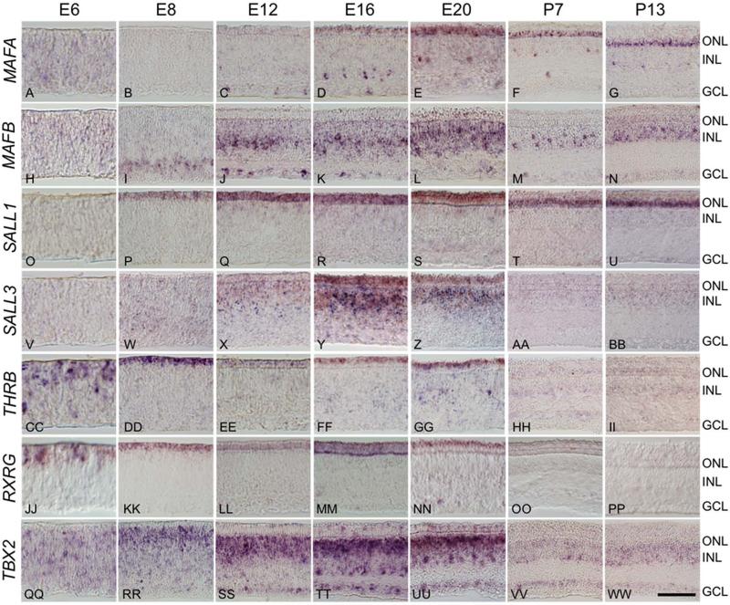
Differentially expressed transcription factors have distinct spatiotemporal expression patterns. In situ hybridization across a developmental time course was used to assess the spatiotemporal expression patterns of a subset of differentially expressed transcription factors. Time points included embryonic days 6 (E6), E8, E12, E16, E20 and post-hatch days 7 (P7) and P13. A-G: Maf family member MAFA localizes to photoreceptors and small population of cells at the inner edge of the INL and GCL beginning at E12 and persisting through P13. H-N: Maf family member MAFB localizes to a subset of photoreceptors at E20, and to a subset of putative amacrine and ganglion cells beginning at E8 and persisting in the adult. O-U: Spalt family member SALL1 expression is largely restricted to the ONL, with faint staining at the outer border of the INL, beginning at E8 and persisting through P13, while SALL3 localizes strongly to the both the ONL and INL in a temporally restricted pattern from E12-E20 (V-BB). CC-PP: Thyroid hormone receptor β2 (THRB) and retinoid × receptor γ (RXRG) are both present as early as E6, persist through E20 and are restricted to the ONL. QQ-WW: T-box transcription factor TBX2 is found in a subset of photoreceptors at E12 through E20, and is widely expressed in the INL and GCL throughout development. ONL = outer nuclear layer, INL = inner nuclear layer, GCL = ganglion cell layer. Scale bar = 50 μm.

