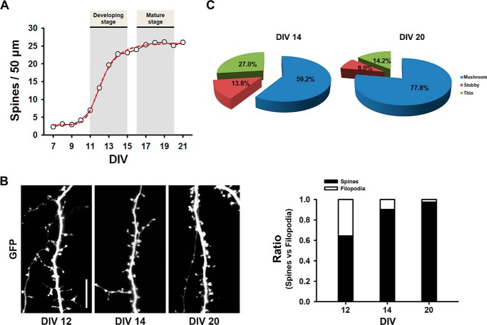
FIGURE 1.
Developmental changes of dendritic spine formation in rat primary cultured hippocampal neurons. A, cultured neurons were transfected with GFP at DIV 7 and then fixed at the indicated DIV. Spine density rises during DIV 11–15, and then the levels persist. Data were collected from three coverslips with each having two dendrites from three neurons at the indicated DIV. B, representative images for spines at the indicated DIV and a ratio graph of spines versus filopodia are depicted. C, pie graphs illustrating the changes in the portion of different morphological types of spine protrusions in the neurons expressing GFP depending on developmental stages.

