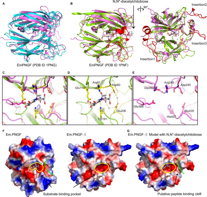
FIGURE 6.
Substrate-binding region and active site of PNGase F-II. A, superimposition of the PNGF domain of PNGase F-II and EmPNG (Protein Data Bank code 1PNG). The PNGF domain of PNGF-II is colored violet, and EmPNG is colored cyan. B, superimposition of the PNGF domain of PNGase F-II and EmPNG with product (N,N′-diacetylchitobiose) (Protein Data Bank code 1PNF). The PNGF domain of PNGF-II is colored violet, and EmPNG is colored aquamarine. The three insertions in PNGF-II are colored red. C, superimposition of the glycan-binding sites of PNGase F and PNGase F-II. D and E, glycan-binding sites of PNGase F and F-II, respectively. F, electrostatic surface potential diagrams of the glycan-binding grooves. The grooves are marked by yellow lines. G, PNGase F-II with an N,N′-diacetylchitobiose molecule modeled in the glycan-binding groove. The putative peptide-binding cleft is marked by green lines.

