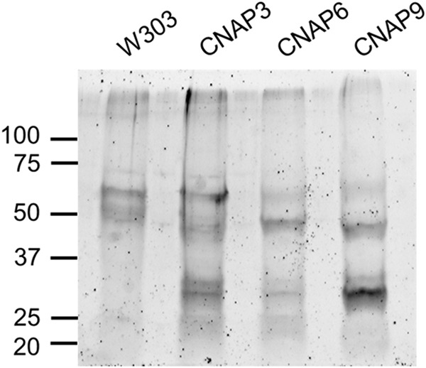
FIGURE 5.

SYPRO Ruby staining for total protein reveals unique bands in CNAP tagged eluates. For each strain designated, aliquots of the anti-PC eluates (20% of E1 and 20% of anti-PC eluate two (E2)) were combined and dried with a SpeedVac at 60 °C for 2 h. Samples were resuspended in 42 μl of SDS sample buffer and separated with an 8–16% Criterion SDS-PAGE gel (Bio-Rad). Following separation proteins were fixed, and the gel was stained overnight with a 1:1 mixture of fresh and used SYPRO Ruby (Life Technologies). The gel was subsequently washed and visualized with an FX Pro Plus Molecular Imager (Bio-Rad) at 532-nm excitation, and emission was measured with a 555-nm long pass filter. The ladder denotes protein masses in kDa.
