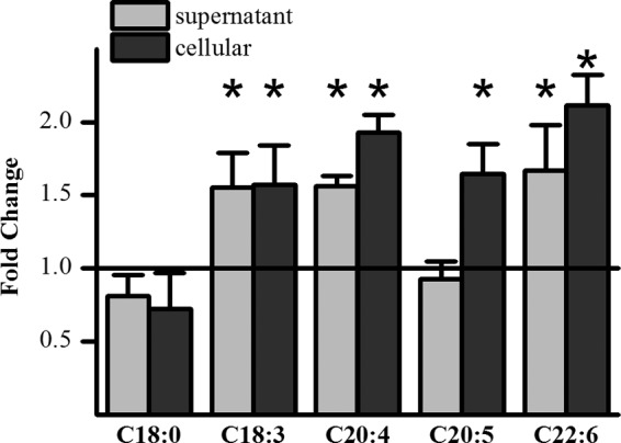
FIGURE 1.

Effect of PUFAs on EC-3′-Glc formation in HepG2 cells. Ratio of EC-3′-Glc concentration detected in the supernatant and the cell lysate of fatty acid-pretreated cells (50 μm, 5 days) compared with the concentration detected in vehicle only-treated cells. Cells were incubated with 200 μm (−)-epicatechin for 2 h. *, p ≤ 0.05; n = 6.
