FIGURE 1.
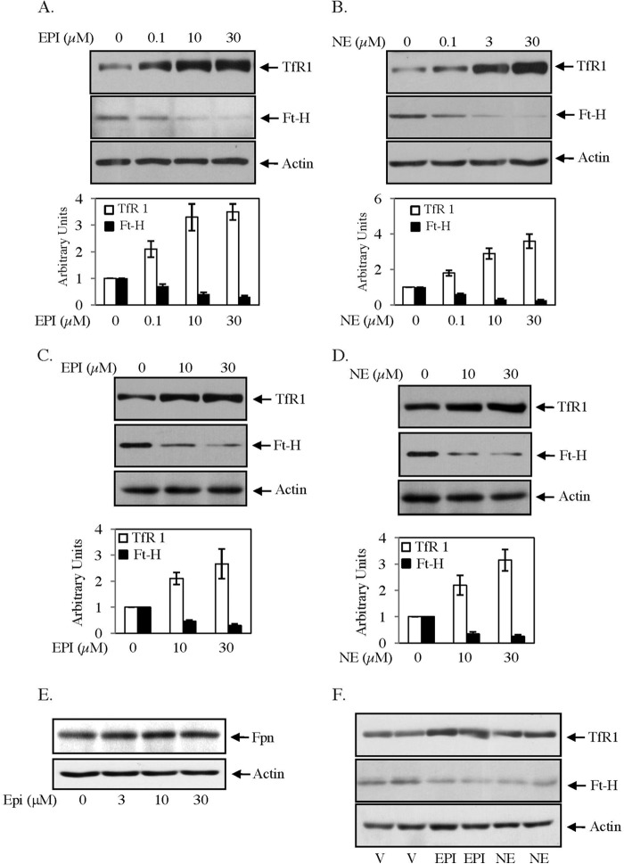
EPI/NE regulates TfR1 and Ft-H expressions in liver and muscle cells. A and B, expressions of TfR1 (upper panel), Ft-H (middle panel), and actin (lower panel) were detected by Western blot analyses in EPI-treated (A) (0–30 μm, 16 h) and NE-treated (B) (0–30 μm, 16 h) HepG2 cell lysates. Bottom panels are quantifications of the Western blot analyses of three independent experiments. Error bars indicate S.D. C and D, similarly, TfR1 (upper panel), Ft-H (middle panel), and actin (lower panel) expressions were analyzed by Western blotting in C2C12 cells stimulated for 16 h with EPI (0–30 μm) (C) and NE (0–30 μm) (D). Bottom panels are quantifications of Western blot analyses of three independent experiments, and error bars indicate S.D. E, HepG2 cells were treated with EPI (0–30 μm) for 16 h, and Western blot analysis was performed using Fpn (upper panel) and actin (lower panel) antibody. The result represents one of the three independent experiments. F, TfR1 (upper panel), Ft-H (middle panel), and actin (lower panel) expressions were detected by Western blot analyses in liver homogenate isolated from vehicle (V)-, EPI-, and NE-injected mice (6 h). Each lane represents homogeneous mixtures isolated from three animals. At least three independent experiments were performed for each group.

