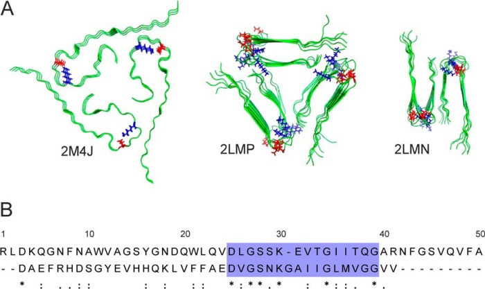
FIGURE 1.
Aβ(1–40) fibrillar structures and sequential similarities with medin. A, structural models of Aβ(1–40) (viewed down the fibril axis) based on solid-state NMR restraints, taken from the Protein Data Bank files indicated. Asp23 and Lys28 are highlighted in red and blue, respectively. B, sequence alignment of medin (top) and Aβ(1–40) (bottom) performed using the LAlign server (29).

