
FIGURE 3.
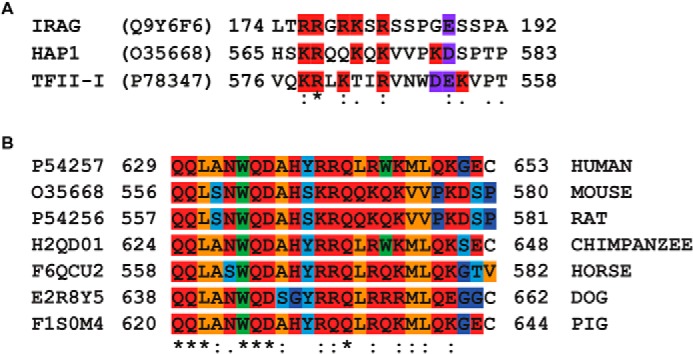
A, alignment of the established IRAG and the reverse TFII-I binding domains to PKG Iβ with the HAP1 region predicted to bind to PKG Iβ. These two sequences show a high degree of similarity regarding the position of the basic and acidic residues. The basic residues are highlighted in red, and the acidic residues are highlighted in purple. B, sequence alignment of the human HAP1 region binding to PKG Iβ with various orthologs in other mammalian species. : and . indicate similarity, and * indicates identity. The residues are color-coded as follows: orange, aliphatic; red, polar; light blue, Ser/Thr/Tyr; green, bulky; and blue, Gly/Pro.