FIGURE 2.
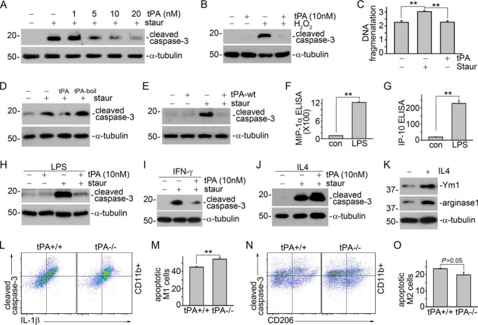
tPA protects M1 macrophage against apoptosis. A, J774 macrophages were treated with 100 nm staurosporine with or without tPA (1, 5, 10, and 20 nm) for 4 h, followed by Western blot for cleaved caspase-3 and α-tubulin. B, J774 cells were treated with 500 μm H2O2 plus 10 nm tPA or vehicle for 16 h, followed by Western blot for cleaved caspase-3 and α-tubulin. C, J774 cells were treated with 100 nm staurosporine with or without 10 nm tPA for 4 h, followed by DNA fragmentation assay. **, p < 0.01, n = 3. D, J774 cells were treated with 100 nm staurosporine with or without 10 nm heat-inactivated tPA (tPA-boil) for 4 h, followed by Western blot for cleaved caspase-3 and α-tubulin. E, J774 cells were treated with 100 nm staurosporine with or without 10 nm wild-type tPA (tPA-wt) for 4 h, followed by Western blot for cleaved caspase-3 and α-tubulin. The supernatants of J774 treated with 1 μg/ml of LPS overnight were subjected to ELISA for MIP-1α (F) and IP-10 (G). **, p < 0.01, n = 5–6. J774 cells were treated with 1 μg/ml of LPS (H), 10 ng/ml of IFN-γ (I), or 100 ng/ml of IL-4 (J) overnight, followed by incubation with 100 nm staurosporine with or without 10 nm tPA for 4 h. Apoptosis was assessed by Western blot for cleaved caspase-3. K, Western blot for Ym1, arginase-1, and α-tubulin in J774 cells treated with 100 ng/ml of IL-4 overnight. Single cell suspensions prepared from whole obstructed kidneys from tPA WT and KO mice were subjected to flow cytometry analyses to quantify cleaved caspse-3-positive IL-1β-expressing (L and M) or CD206-expressing (N and O) CD11b+ macrophages. L and N, representative pictures of flow cytometry analyses. M and O, quantitative illustrations. **, p < 0.01, n = 4 mice per group.

