FIGURE 5.
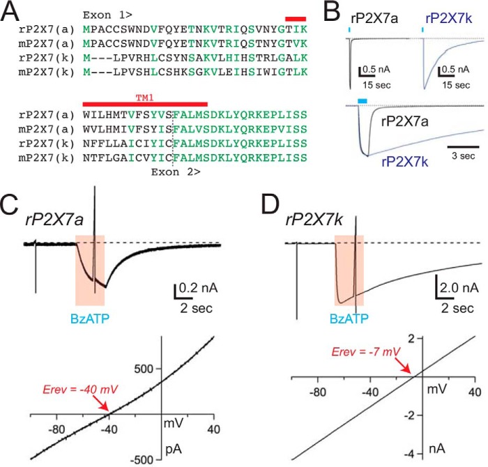
The rP2X7kR is constitutively dilated. A, sequence alignment of the N terminus and TM1 of rat and mouse P2X7a and P2X7k receptors. Identical and similar residues are shown in green. TM1 is marked with a red bar. B, the top two traces show the time courses of the membrane currents gated by 100 μm BzATP in voltage-clamped HEK293 cells (holding voltage = −60 mV) expressing either rP2X7aRs (left) or rP2X7kRs (right). The rP2X7kR often took several minutes to fully deactivate upon washout of agonist. In the bottom trace, normalized rP2X7aR and rP2X7kR currents are overlaid to show the difference in deactivation times. C and D, voltage ramps (140 mV, 200 ms) from a holding voltage of −100 mV were applied to HEK293 cells expressing either rP2X7aRs (panel C) or rP2X7kRs (panel D) before and during applications of 30 μm BzATP. Currents through the rP2X7aR were reversed at membrane potentials (bottom left trace) that were significantly more negative than the currents through the rP2X7kR (bottom right trace). These data support the hypothesis that the pore of the rP2X7kR is constitutively dilated. Similar results were found using the mouse splice variants.

