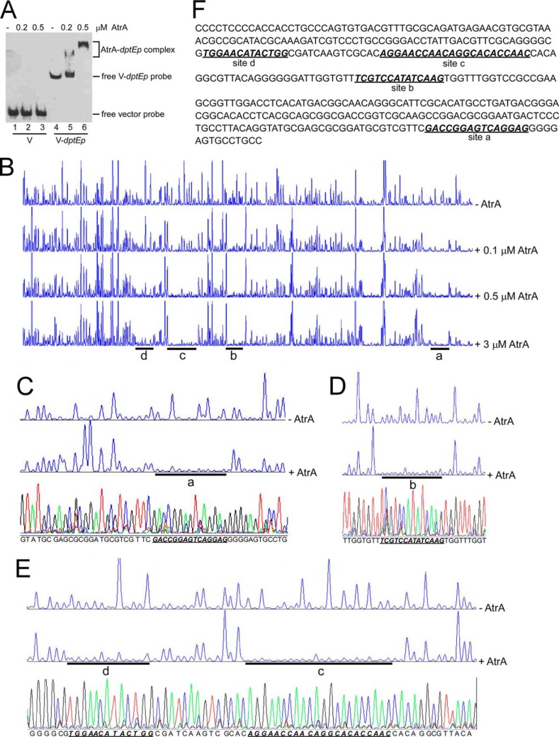
FIGURE 2.
Binding of AtrA to the dptE promoter (dptEp). A, EMSA of AtrA binding to dptEp. Biotin-labeled dptEp (V-dptEp) was used as a probe, and biotin-labeled void vector (V) was the negative control. B, DNase I footprinting assays of AtrA binding sites on dptEp. FAM-labeled dptEp was used as a probe with gradient concentrations of AtrA. The protected regions were labeled as site a, b, c, and d, respectively. C–E, binding sequence determination of AtrA on dptEp by DNase I footprinting assays for site a (C), site b (D), and sites c and d (E). F, the binding sequences of AtrA on dptEp as determined by DNase I footprinting assays. The four binding sites are shown in bold italics and are underlined.

