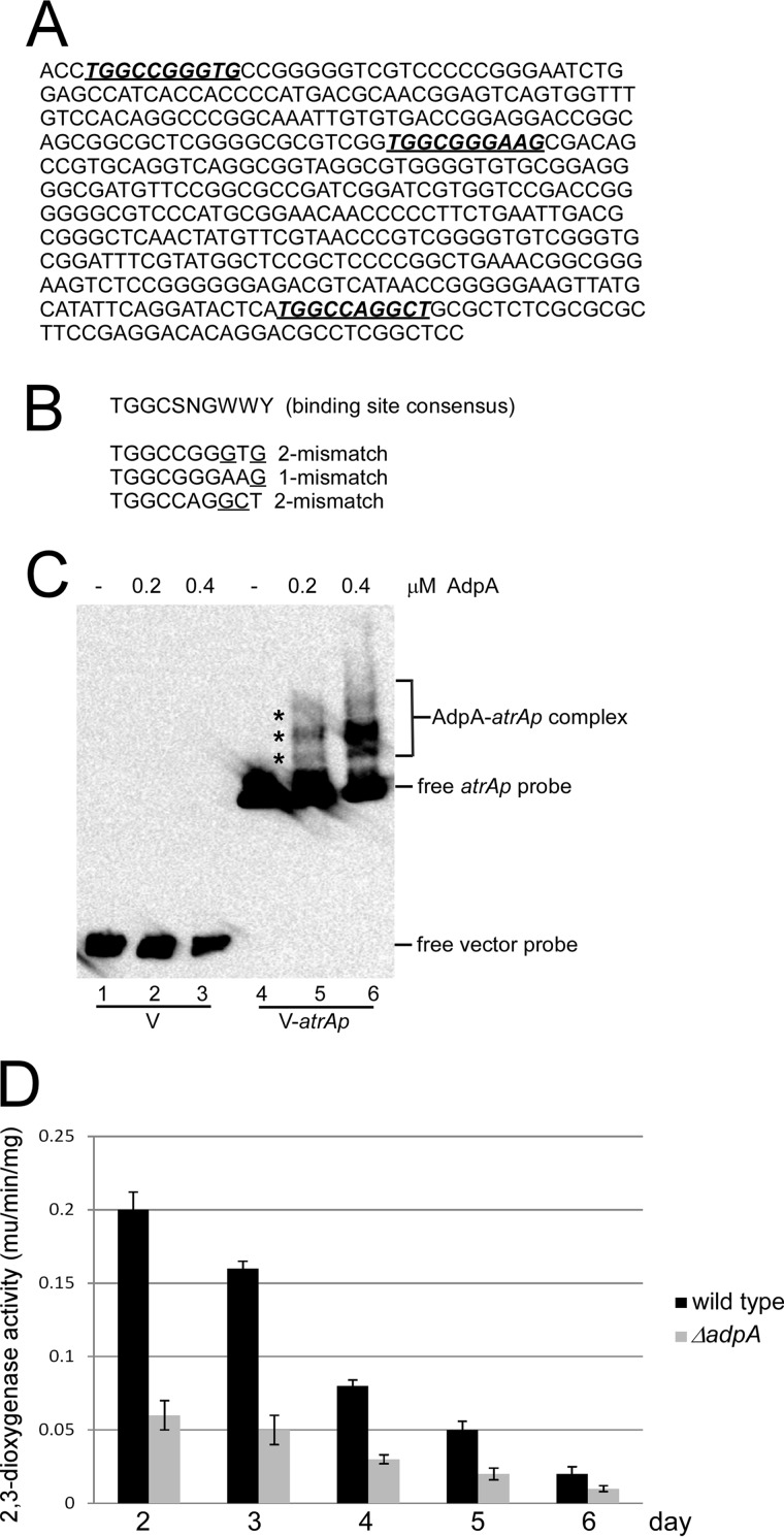
FIGURE 5.
Binding of AdpA to atrA promoter (atrAp). A, DNA sequence of atrAp. The deduced AdpA binding sites are shown in bold italics and underlined. B, alignment of deduced AdpA binding sites on atrAp with the binding consensus. One or two mismatched nucleotides are indicated. C, EMSA of AdpA binding to atrAp. Biotin-labeled atrAp (V-atrAp) was used as a probe, and biotin-labeled void vector (V) was the negative control. D, AdpA positively regulates atrAp activity. The wild type and the ΔadpA mutant were transformed with pIPP1-atrAp (atrAp-xylE) and cultured in YEME medium. Cells were collected and disrupted by sonication every day, and 2,3-dioxygenase activity was measured. Triplicate independent experiments were demonstrated, and S.D. bars are shown.

