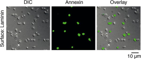
Fig. 2.

Laminin-bound platelets expose phosphatidylserine (PS). Washed human platelets were exposed to a surface of immobilized laminin. Bound platelets were incubated with a solution of fluorescently-labeled annexin V to visualize PS exposure. Images were recorded using DIC and fluorescence microscopy. Corresponding brightfield and fluorescent images are shown alone and in the overlay.
