
Figure 1.
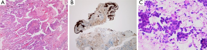
Pathology sample types of lung cancer. (A) Surgical resection specimen. Pathology work-up, paraformaldehyde fixation, paraffin embedding, HE staining (20×); (B) transbronchial biopsy. Paraformaldehyde fixation, whole sample embedding to paraffin, immunohistochemistry for EGFR (40×). Note the normal surface epithelium and a minimal amount of stromal tumor cell clusters; (C) cytology smear. Paraformaldehyde fixation, HE staining (100×). Note the tumor cell rich area.