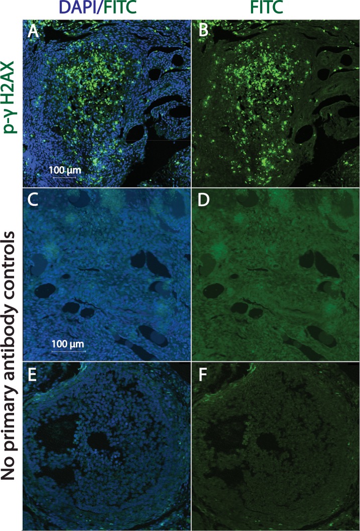
FIG. 5.
DXR-induced formation of phospho-γH2AX foci within granulosa cells of corpus luteum of marmoset ovarian tissue. Ovarian sections were stained for p-γH2AX (green) and nuclei (blue, DAPI). Fluorescent images A and B illustrate corpus luteum exhibiting positive p-γH2AX staining. Photomicrographs C and D illustrate control ovarian tissue with no primary antibody added (no antral follicles seen). Photomicrographs E and F illustrate control ovarian tissue showing antral follicles with no primary antibody added. Bar = 100 μm.

