
Figure 1.
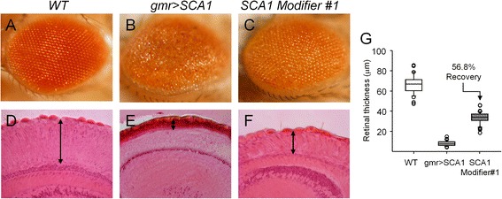
Histological method to analyze Drosophila eye degeneration phenotypes. External pictures of fly eyes surface (A-C) and histological measurement of retinal thickness (D-F). Images in a column correspond to the same genotype. (G) Retinal thickness quantification in μm, resulting in a significant recovery from the degenerated genotype for the gmr > SCA1 Modifier#1 (n = 34-46 sections/genotype).