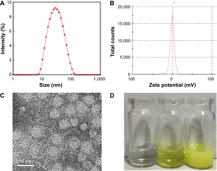
Figure 2.
Characterizations of the Qu-M.
Notes: (A) Size distribution of Qu-M, (B) zeta potential of Qu-M, (C) TEM image of Qu-M, (D) PBS solution, Qu-M in PBS solution, and free quercetin in PBS solution (from left to right).
Abbreviations: Qu-M, quercetin-loaded MPEG–PCL nanomicelles; TEM, transmission electron microscope; PBS, phosphate-buffered saline; MPEG–PCL, monomethoxy poly(ethylene glycol)–poly(ε-caprolactone).

