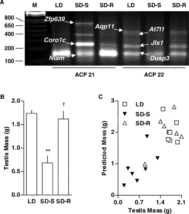
Figure 1. Photoperiodic regulation of testicular expression and functionality.
(A) Differential display PCR was used to assess changes in testicular transcript levels of hamsters exposed to long days (LD: 14 h light and 10 h dark for 16 weeks, n = 7), short days during the photosensitive period (SD-S: 10 h light and 14 h dark for 16 weeks, n = 7), or short days during the photorefractory period (SD-R: 10 h light and 14 h dark for 22 weeks, n = 6).Differentially represented cDNAs were extracted from the agarose gel, cloned into the TOPO-TA cloning vector, and sequenced. Among the genes whose roles in testicular function have not been established, we identified aquaporin-11 (Aqp11), ataxin-7-like protein-1 (At7l1), coronin actin-binding protein-1c, dual specificity phosphatase-3 (Dusp3), nuclear interactor of Arf and Mdm2 (Niam), a cDNA of undetermined identity (Jls1), and zinc finger protein-639 (Zfp639). (B) Relative to LD exposure, SD-S exposure decreased testis weight by 61%. Relative to SD-S exposure, SD-R exposure increased testis weight by 140%. Means (± SEM) are shown. For LD vs SD-S comparison, **p < 0.01. For SD-S vs SD-R comparison, †p < 0.01. (C) Multivariate regression analysis, across the three photoperiodic treatments, revealed that Aqp11 gene expression had the highest correlation with testis weight.

