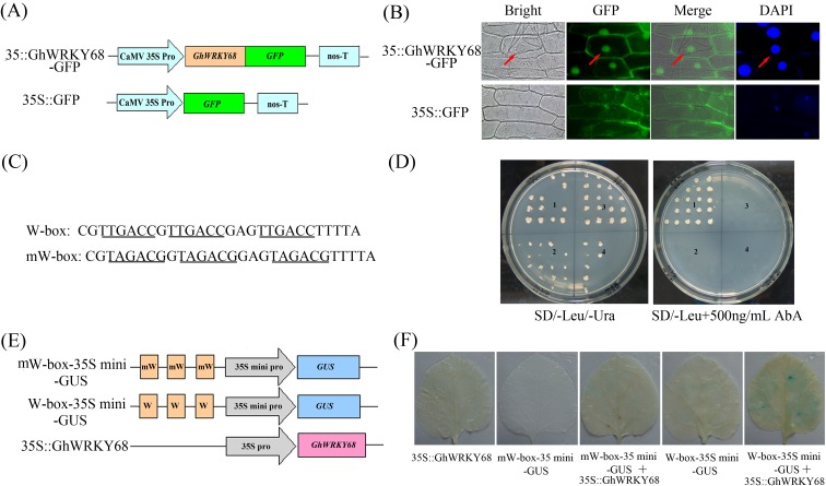
Fig 2. Characterisation of GhWRKY68 as a transcription factor.
(A) Schematic diagram of the 35S-GhWRKY68::GFP fusion protein construct and the control 35S-GFP construct. GFP was fused in frame to the C terminus of GhWRKY68. (B) Transient expression of the 35S-GhWRKY68::GFP and 35S-GFP constructs in onion epidermal cells 24 h after particle bombardment. Green fluorescence was observed using a fluorescence microscope, and the nuclei of the onion cells were visualised by DAPI staining. (C) Sequence of the triple tandem repeats of the W-box and mW-box. (D) Transactivation analysis of GhWRKY68 with the yeast one-hybrid assay using the 3×W-box or mW-box as bait. Yeast cells carrying pGAD-GhWRKY68 or pGAD7 were grown on SD/-Leu/-Ura or SD/-Leu containing 500 ng/ml AbA. 1, pAbAi-W-box/pGAD-GhWRKY68, 2, pAbAi-W-box/pGAD7, 3, pAbAi-mW-box/pGAD-GhWRKY68, and 4 pAbAi-mW-box/pGAD7. (E) Schematic diagram of the reporter and effector constructs used for co-transfection. (F) Histochemical analysis of co-transfected N. benthamiana leaves. Fully expanded leaves from 8-week-old N. benthamiana were agro-infiltrated with the indicated reporter and effector at an OD600 of 0.6. GUS staining was performed 3 days after the transformation.

