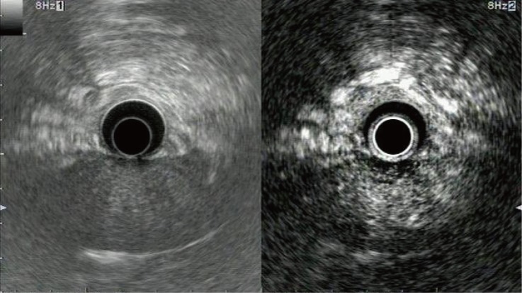
Fig 2. Representative example of focal pancreatitis with hyperenhancement.
Conventional endoscopic ultrasonography (left) shows a slightly hypoechoic area without a clear margin at the pancreas head. Contrast-enhanced harmonic endoscopic ultrasonography (right) indicates that enhancement in this area is higher than in the surrounding tissue, and a margin is clearly visible.

