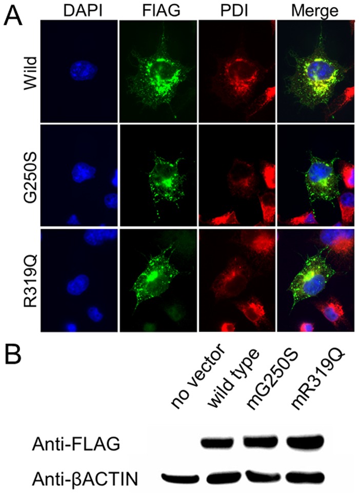
Fig 4. In vitro functional evaluation of SNVs effects.
(A) Immunofluorescence staining of COS1 cells transfected with SNV-harboring CLCN6 variants. Protein disulfide isomerase (PDI) is used as marker of the endoplasmic reticulum (ER). FLAG-tagged CLCN6 is merged with PDI, indicating CLCN6 localization in the ER. (B) Western blotting analysis of cell lysates shows no difference in CLCN6 expression.

