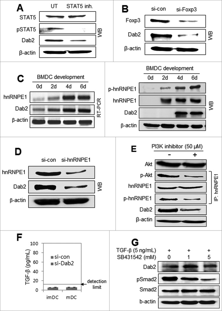
Figure 2 (See previous page).
Dab2 expression in DCs requires STAT5 signaling, Foxp3 expression, and hnRNPE1 activation but has no association with TGF-β signaling. (A) Mouse (C57BL/6) BMDCs were treated with 100 μM STAT5 inhibitor for 4 h before harvest. Harvested cells were subjected to Western blot analysis of STAT5 and Dab2 expression with STAT5, phospho-STAT5, and Dab2 antibodies. (B) DC precursor cells on day 4 during BMDC development were transfected with control and Foxp3 siRNAs and then harvested after 48 h. Dab2 expression was assessed by Western blot with Dab2 and Foxp3 antibodies. (C) mRNA (left) and protein (right) expressions of hnRNPE1 and Dab2, together with hnRNPE1 phosphorylation, were assessed during BMDC ←development by RT-PCR and Western blot analysis. (D) BMDC precursor cells on day 4 were transfected with si-con and si-hnRNPE1 and harvested after 48 h. Dab2 and hnRNPE1 expression was assessed by Western blot with Dab2 and hnRNPE1 antibodies. (E) BMDCs were treated with 50 μM PI3K inhibitor (Calbiochem) for 20 min before harvest and subjected to Western blot analysis of Akt and Dab2. hnRNPE1 was immunoprecipitated from BMDC lysates with anti-hnRNPE1 antibody, followed by immunoblot analysis with phospho-hnRNPE1 (phospho-serine) and phospho-Akt antibodies. (F) BMDC precursor cells on day 4 were transfected with Dab2-specific siRNAs (si-Dab2) or control siRNA (si-con). After 48 h, the level of TGF-β in culture supernatant was examined using TGF-β ELISA kit (BioLegend). (G) BMDCs were treated or untreated with 5 ng/mL TGF-β for 24 h in the presence or absence of SB431524 (TGFβRI). Dab2 and Smad2 expressions, and phospho-Smad2 were assessed by Western blot.

