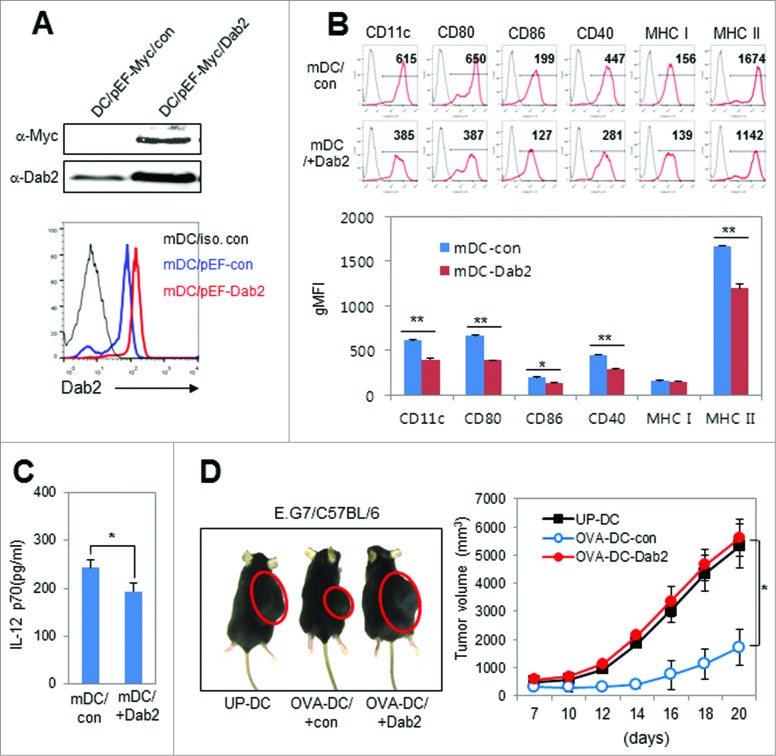
Figure 7.
Dab2-overexpression impaired the efficacy of the DC vaccine for tumor immunotherapy. (A) Dab2 expression plasmid (pEF-Myc/Dab2) and control vector (pEF-Myc/con) were transfected into DC precursor cells on day 4 during BMDC development. The transfected cells were harvested after 48 h, and matured with LPS (100 ng/mL) for 24 h. Dab2 expression in the transformed mDCs was assessed by Western blot (upper) and FACS analysis (lower) using anti-Myc and anti-Dab2 antibodies. (B) The surface phenotypes of Dab2 transfected mDCs were assessed by flow cytometry, and the gMFI value of each DC surface marker from three independent experiments is shown as mean ± SD of nine samples. *p < 0.05, **p < 0.01 compared with control vector-transfected DCs, Student's t-test. (C) IL-12 levels were assessed by ELISA of the culture supernatant of Dab2-transfected DCs and are shown as mean ± SD of nine samples. *p < 0.05 compared with control vector transfected DCs, Student's t-test. (D) C57BL/6 mice were inoculated s.c. with E.G7 tumor cells (5 × 10⁵) in the right flank, and then immunized on day 3 and day 10 with 1 × 10⁶ Dab2-expressing mDCs (OVA-DC-Dab2) or control vector-transfected mDCs (OVA-DC-con), which were derived from C57BL/6 BM cells and pulsed with OVA peptides (OVA257–264 and OVA323–339). Representative images of E.G7 tumors are shown on day 20 after DC vaccination (left). Tumor growth was monitored every 2–3 d and presented as mean ± SD of four mice from each of two experiments. *p < 0.05, Student's t-test.

