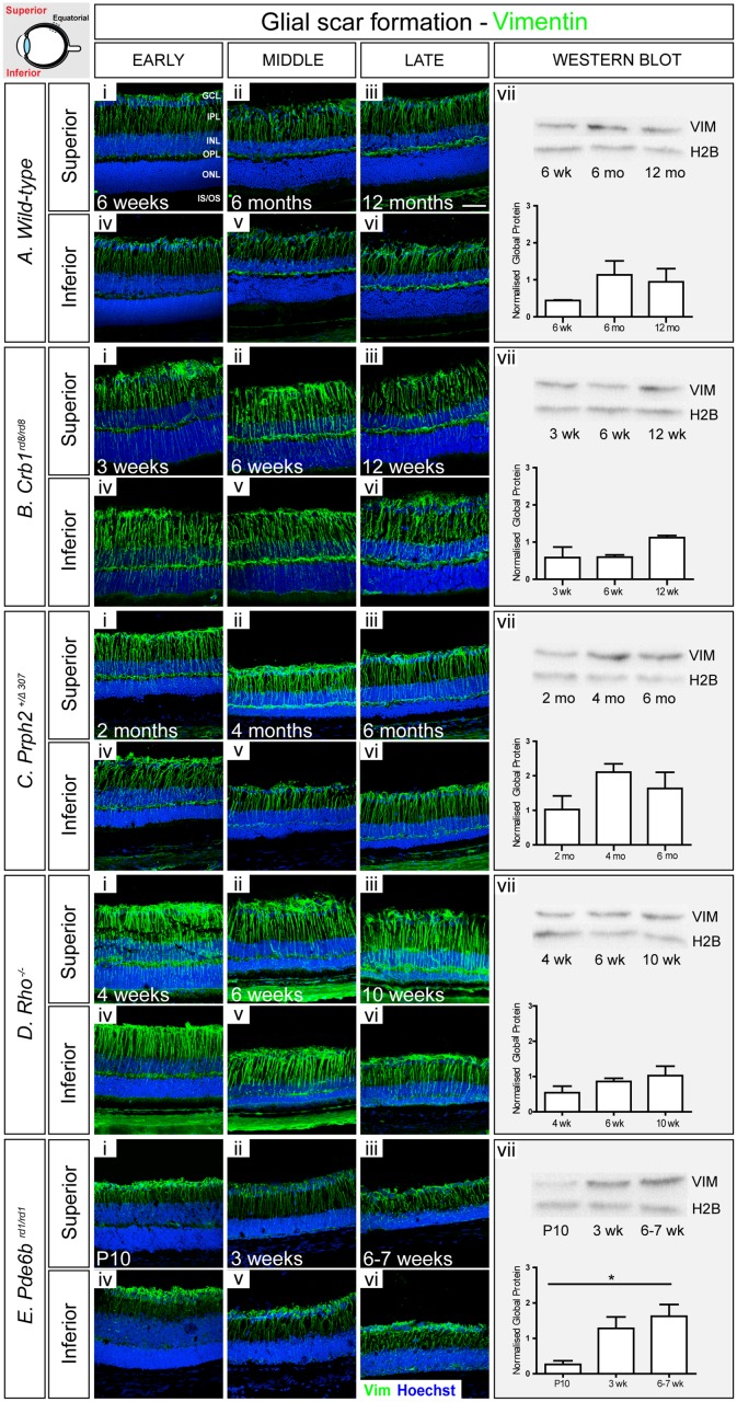
Fig 3. Vimentin increases with disease progression.
A. In wild-type retinae, vimentin+ve fibers extended throughout the retina at all examined ages (i-vi). Staining at the level of whole retina increased slightly with age (vii). B. In Crb1 rd8/rd8 model, the amount of vimentin was higher, particularly in the outer retina, compared to wild-type (i-vi). A small increase in global level was observed at the latest stage examined (vii). C. Prph2 +/Δ307 animals showed a similar pattern of vimentin to wild-type, with additional glial processes extending throughout the ONL at the early stage (i-vi), although no significant increase was observed, as determined by Western Blot (vii). D. In Rho -/- mice, vimentin+ve processes were numerous and more prominent than in wild-type retinae (i-vi), but no significant change in global level was observed (vii). E. In Pde6b rd1/rd1, robust staining for vimentin was seen throughout (i-vi) and global levels increased significantly with degeneration (vii). Cryosections were immunostained for vimentin (green) and counterstained with nuclei marker Hoechst 33342 (blue). Scale bar, 50 μm. Semiquantitative assessments of vimentin in whole neural retinae were determined by Western blot. Vimentin was normalized against H2B. Statistical significance was assessed with a one-way ANOVA test with Tukey’s correction; *P < 0.05, **P < 0.01, and ***P < 0.001. GCL, ganglion cell layer; IPL, inner plexiform layer; INL, inner nuclear layer; OPL, outer plexiform layer; ONL, outer nuclear layer; IS/OS, photoreceptor inner/outer segment region.

