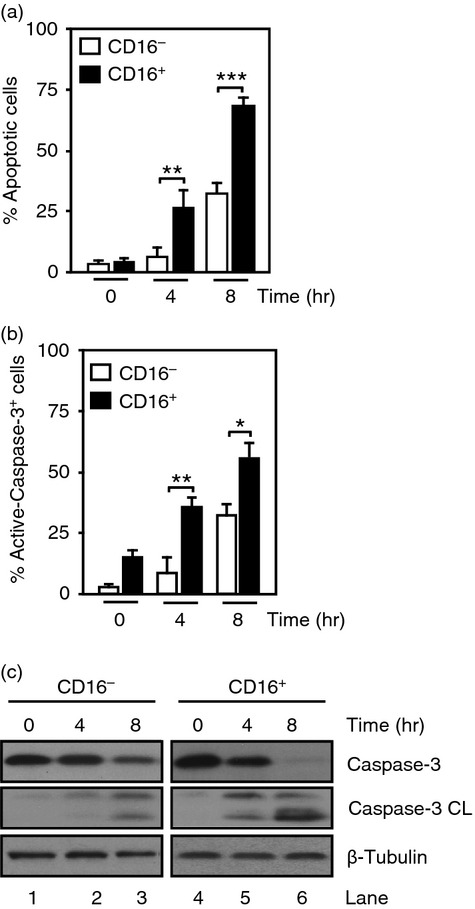
Figure 1.

Spontaneous apoptosis in monocyte subsets. Purified human CD16− and CD16+ monocytes were cultured in serum-free media for 4 and 8 hr. (a) Percentage of apoptotic cells was assessed by Annexin V/7-AAD staining. (b) Percentage of cells stained with an anti-active-caspase-3-phycoerythrin antibody. Data represent mean ± SEM (n = 3, *P < 0·05, **P < 0·01, ***P < 0·001). (c) Immunoblots from the samples used in (a) probed with anti-inactive-caspase-3 (Casp-3) or anti-active-caspase-3 (Casp-3 CL) antibodies. β-Tubulin expression was used as loading control. Data are representative of three experiments.
