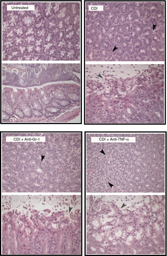
Figure 4.

Photomicrographs of representative haematoxylin & eosin (H&E) -stained colonic sections from Untreated, Clostridium difficile-infected, C. difficile-infected and anti-Gr-1-treated, C. difficile-infected and anti- tumour necrosis factor-α (TNF-α)-treated animals (day 4). Cross-sections of colonic crypts (upper images) and longitudinal sections of the epithelial–luminal interface (lower images) are shown for each treatment. Black arrowheads indicate infiltrating inflammatory cells, while grey arrowheads indicate areas of epithelial damage. All animals were treated as outlined in Fig.1. Total magnification for all images is 400 ×. CDI = C. difficile infected.
