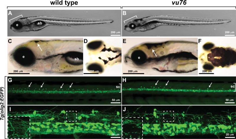
Figure 1. The vu76 mutation causes defects in pigment deposition and oligodendrocyte development.

Images of living wild-type (A) and vu76 mutant (B) larvae at 6 dpf. The mutant has a smaller brain (bracket) and eye (e) than wild type and lacks a swim bladder (sb). Lateral (C, E) and dorsal (D, F) views of 5 dpf wild-type and vu76 mutant larvae to show pigment patterns. The dark pigment of the mutant is more broadly distributed than that of the wild type. Low (G, H) and high (I, J) magnification images of Tg(olig2:EGFP) reporter gene expression in wild-type and mutant larvae. Images are focused on the trunk spinal cord (sc, brackets) with dorsal up. In G and H dorsally migrated OPCs appear as green dots (arrows). Fewer dorsal OPCs are apparent in the vu76 mutant larva than in the wild-type larva. At 6 dpf OPC and oligodendrocyte processes form a dense meshwork (I, boxed area magnified as inset). Oligodendrocyte lineage cell processes are evident in the mutant larva, but the density of processes is less than in wild-type (J, boxed area magnified as inset).
