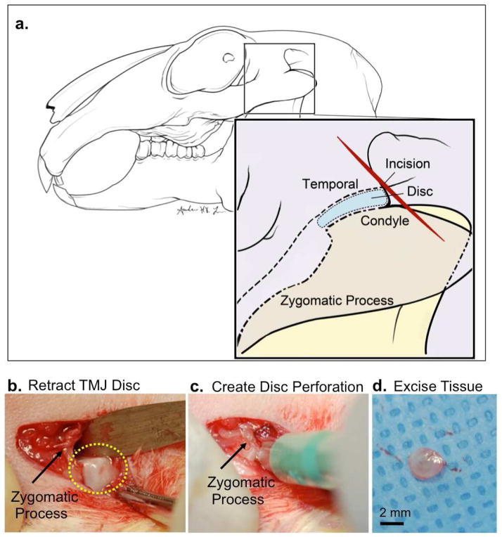
Figure 1. Rabbit TMJ injury model.
New Zealand white rabbits (N=9) underwent unilateral TMJ disc perforation surgery on the left TMJ. Sham surgery was performed on the right contralateral TMJ within the same rabbit and served as a comparison control. (a) Diagram illustrating New Zealand white rabbit TMJ anatomy. The disc is localized behind the zygomatic process. An oblique incision was made superior to the zygomatic process. (b) The TMJ superior joint space was accessed and forceps were used to retract the disc (yellow dashed lines) posterior to the zygomatic process (black arrow). (c) A punch biopsy was used to create a 2.5 mm perforation in the anterior lateral TMJ disc. (d) A 2.5 mm tissue was excised from the TMJ disc and the incision was sutured. Scale bar = 2mm.

