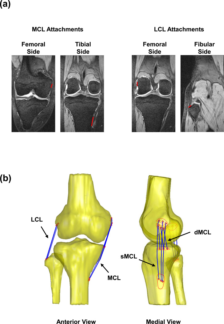
Figure 1.
(a) Coronal plane MR images of the knee were digitized and used to create the femoral and tibial attachment areas of the MCL and LCL. (b) Anterior view (left) and medial view (right) of a 3D knee model created using Sagittal plane MR images. The knee model includes the 3D surface of the femur, tibia and fibula (yellow), and the attachments of the medial and lateral collateral ligaments. Each Ligament was evenly divided into 3 portions (blue curves), by connecting the centroids of corresponding segments (red circles).

