
Fig 1. Representative photomicrograph of a coronal section of the rat brain at the level of the NTS illustrating the microinjection site (encircled area) for the behavioral experiments (right panel) and a schematic representation of the NTS from the rat atlas (Paxinos and Watson, 1998) (left panel).
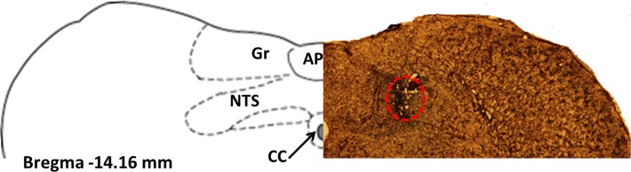
Area postrema (AP), central canal (cc), gracile nucleus (Gr).