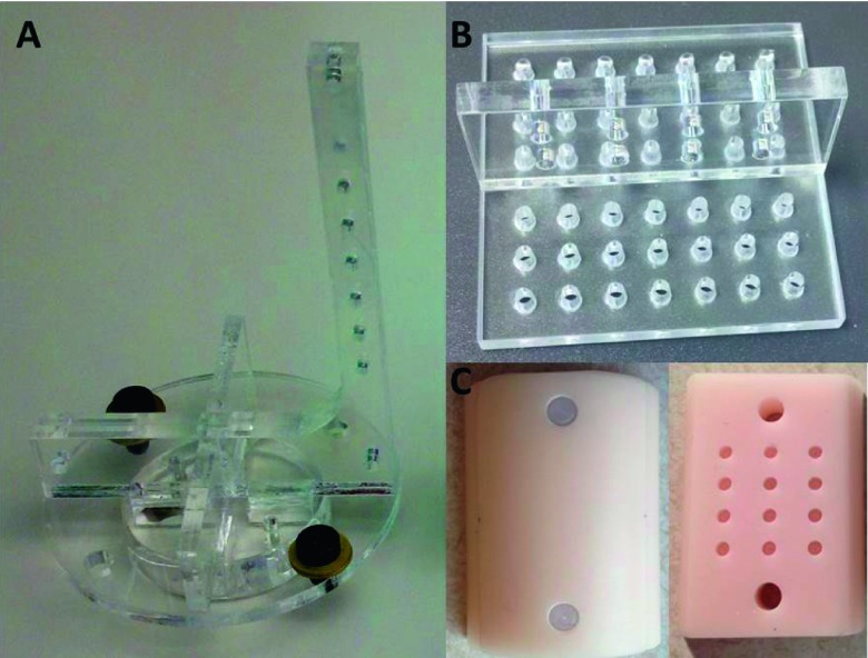
FIG. 2.
Phantoms used in calibration. (A): The seven-bead phantom for CBCT geometrical calibration. (B): The acrylic phantom with holes for the geometrical calibration of the entire imaging system. (C): The half cylinder tissue-simulating phantom for validation of the absolute emittance and BLT reconstruction. The left piece is the cover and the right is the bottom containing 12 holes for placing luminescent sources.

