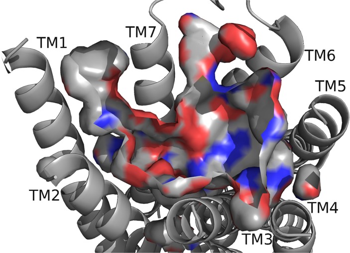
Fig 3. DrmMS-R2 model.
The receptor backbone, binding pocket, with the exposed atoms are highlighted, and TMs are shown; refer to Fig. 2 for notation. The deepest portion of the pocket was in the region near TM5. DrmMS-R2 contained a highly polar region between TM6 and TM7 and two hydrophobic and aromatic regions near TM1 and TM5.

