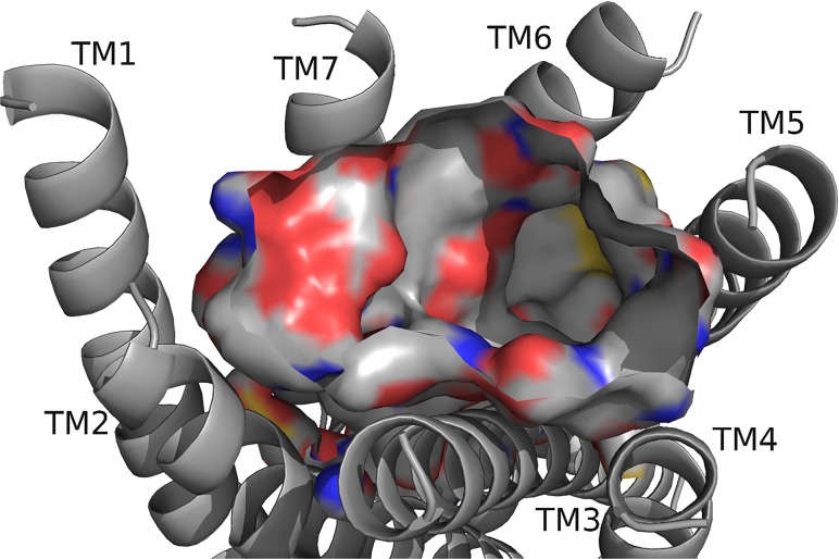
Fig 13. RhpMS-R model.
The RhpMS-R backbone and the exposed atoms at the surface of the binding pocket are highlighted; refer to Fig. 2 for notation, including sulfur represented as yellow. The pocket contained a highly polar region near TM7 and hydrophobic and aromatic regions near TM5 and TM3. Residues on TM1 were inaccessible for ligand interactions and the pocket was deepest between TM5 and TM6.

