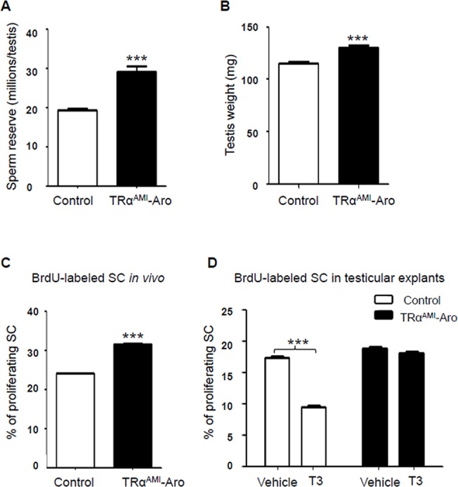
Fig 4. Whole testicular sperm reserve and testis weight in adult TRαAMI-ARO, and percentage of proliferating SC in TRαAMI-ARO at P3 in vivo and in testicular explants using organotypic in vitro cultures with or without exogenous T3.

We observed a significant increase in whole testicular sperm reserve (a) and in testis weight (b) (***P < 0.001; n = 15 for controls and n = 16 for TRαAMI-ARO group). Data are shown as mean ± SEM. Statistical analyses were performed using Student’s t-test. White bars, control; black bars, TRαAMI-ARO (a, b). After BrdU immunohistochemical labeling, BrdU negative and BrdU positive SC were counted and the SC proliferation index was calculated. (c) When BrdU was injected in vivo 3 h before sacrifice, proliferation of Sertoli cells increased in P3 testes of TRαAMI-ARO mice in comparison with the control (***P < 0.001; n = 5 animals for each genotype). (d) When BrdU was added in vitro 3 h before the end of the organotypic cultures of P3 testicular explants, the SC proliferation index significantly decreased in the control in presence of T3 compared with vehicle (***P < 0.001, n = 6). In contrast, T3 had no effect on SC proliferative index in TRαAMI-ARO mice (P > 0.05, n = 5). Data are shown as mean ± SEM. Statistical analyses: two-way ANOVA followed by Bonferroni’s post-hoc test. White bars, control; black bars, TRαAMI-ARO.
