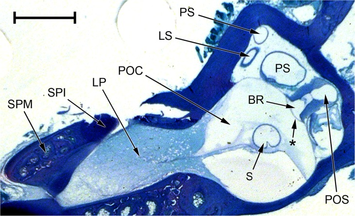
FIG. 11.
Photomicrograph of an oblique section through the inner ear of Xenopus laevis (male specimen). Note that the periotic cistern (POC), the contents of which have picked up only a small amount of stain, is separated from the stapes footplate (SPI and SPM) by the lateral passage (LP), which is filled with diffuse material staining pale blue. The contact membrane of the basilar recess is the thin membrane between the basilar recess (BR) and the periotic sac (POS); the membrane marked with an asterisk between the basilar recess and the POC is Paterson’s (1949, 1960) additional ‘tympanal area’ (see text). Relative to the centre of the picture, dorsal is upwards and away from the viewer, lateral is to the left and towards the viewer. Scale bar, 1 mm. See Fig. 2 caption for full list of abbreviations.

