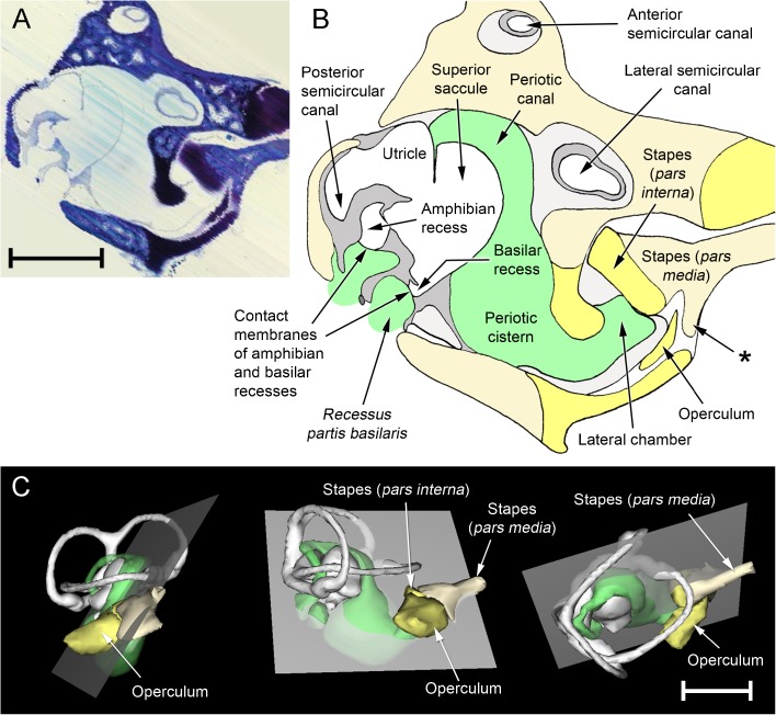
FIG. 12.
Reconstructions created from a stack of serial section images of the ear of Rana pipiens, digitally resectioned in a plane as close as possible to Wever’s (1973) illustration (Fig. 1). See text for details. The extrastapes, tympanic membrane, periotic sac and round window were not within the original sections and are consequently not shown in these reconstructions. A ‘Virtual section’; the faint, diagonal striations indicate the planes of the original section photomicrographs from which this was reconstructed. B Expanded, diagrammatic illustration of the same. Out of the plane of this particular section, the periotic canal is in communication with the periotic fluid which abuts the contact membranes of the amphibian and basilar recesses; these regions are all components of the periotic labyrinth and are hence shaded in green. The asterisk indicates a process of the stapes pars media which articulates with the otic capsule. C WinSurf reconstructions of the right inner ear of R. pipiens from (left) lateral, (middle) posterior and (right) dorsal views, showing the position of the ‘virtual section’ as a grey plane. The stapes and operculum are included in these reconstructions but the internal walls of the otic capsule are not. Both scale bars, 2 mm. Colour code: white = otic labyrinth (endolymph); green = periotic labyrinth (perilymph); dark grey = limbic tissue; lighter grey = looser periotic tissue; cream = bone; yellow = cartilage

