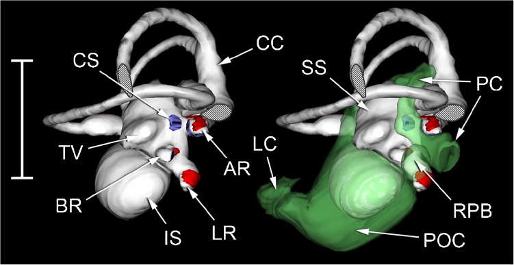
FIG. 3.
WinSurf reconstructions of left inner ear structures of Rana pipiens, seen from an approximately posterior view. The reconstruction on the left shows the otic labyrinth (white), sensory epithelia (red) and contact membranes separating endo- and perilymph (purple). Part of the posterior semicircular canal has been removed to reveal the diverticula of the superior saccule. The reconstruction on the right shows the same, with the periotic labyrinth added in (semitranslucent green). Scale bar, 2.5 mm. See Fig. 2 caption for full list of abbreviations.

