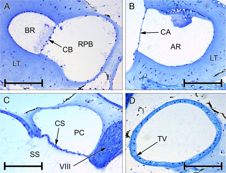
FIG. 5.
Photomicrographs of sections through the inner ear of Rana pipiens. A Basilar recess (BR; endolymph), recessus partis basilaris (RPB; perilymph) and contact membrane of basilar recess (CB) separating the two. B Amphibian recess (AR) and its contact membrane (CA), expanded from Fig. 4A. C Periotic canal (PC) and contact membrane of the saccule (CS), expanded from Fig. 4B. D Tegmentum vasculosum (TV), expanded from Fig. 4A. All scale bars, 200 μm. See Fig. 2 caption for full list of abbreviations.

