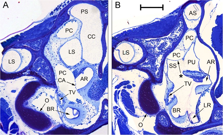
FIG. 7.
Composite photomicrographs of two sections through the inner ear of Eleutherodactylus limbatus. A An approximately transverse plane, through the posterior part of the inner ear. Relative to the centre of the photomicrograph, dorsal is upwards and slightly away from the viewer, lateral is to the left. B An oblique plane (between transverse and sagittal), taken from the ear on the contralateral side to (A) and laterally inverted to aid comparison. Relative to the centre of the photomicrograph, dorsal is upwards, lateral is to the left and away from the viewer. Note in particular the membrane marked with an asterisk, located between the ventral diverticulum of the central part of the periotic canal (PC) and the superior saccular chamber (SS). The membranous labyrinth has pulled away from the otic capsule wall on the right-hand side. Scale bar applies to both (A) and (B) and represents 200 μm. See Fig. 2 caption for full list of abbreviations.

