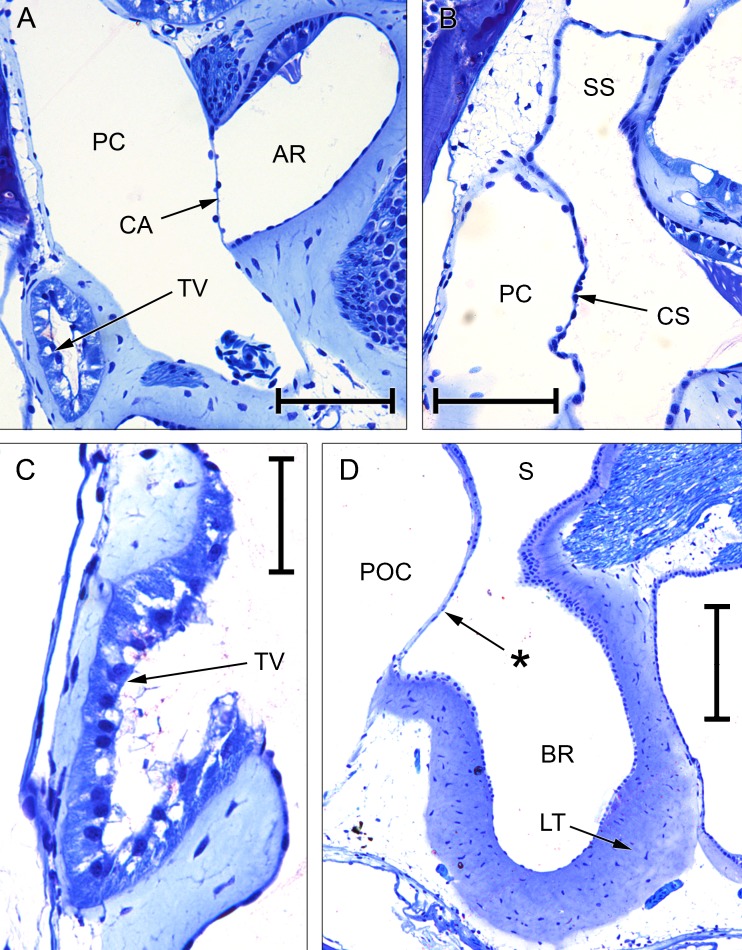
FIG. 8.
Photomicrographs of sections through the inner ears of Eleutherodactylus limbatus and Xenopus laevis. A Tegmentum vasculosum (TV), amphibian recess (AR) and its contact membrane (CA) in E. limbatus; scale bar, 100 μm. B Periotic canal (PC) and the contact membrane of the saccule (CS) in E. limbatus; scale bar, 100 μm. C Tegmentum vasculosum of E. limbatus; scale bar, 50 μm. D Basilar recess (BR) of X. laevis (female specimen); scale bar, 200 μm. The asterisk indicates the additional ‘tympanal area’ identified by Paterson (1949, 1960), located just rostrolateral to the basilar recess (see text). See Fig. 2 caption for full list of abbreviations.

