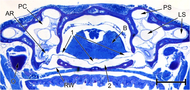
FIG. 9.
Photomicrograph of a transverse section through the head of Eleutherodactylus limbatus, at the level of the posterior half of the otic capsule. The three fluid compartments collectively denoted ‘1’ appear to be separate in this section, but inspection of other sections in the same series suggests that they are actually part of one continuous fluid system extending between the periotic labyrinths of each ear and passing underneath the brain (B). The left-hand arrow marked ‘1’ points to the left periotic sac, which can be seen emerging from the left otic capsule. Within the capsule, it is immediately adjacent to the amphibian recess (AR), from which it is separated by a thin contact membrane. Between fluid space 1 and the base of the skull is a second fluid space (2), which is also continuous across the head and extends between the two round windows (RW). Distortion resulting from shrinkage may have changed the relative sizes of fluid spaces 1 and 2. Scale bar, 0.5 mm. See Fig. 2 caption for full list of abbreviations.

