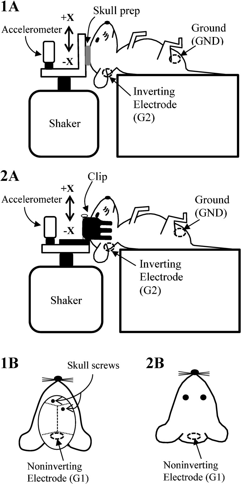
FIG. 5.
Coupling methods for delivering stimulus. The animal’s head was surgically coupled to the aluminum platform using skull screws (1a and 1b) or coupled non-surgically using a non-invasive head clip (2a and 2b). The electrodes for the skull prep method were placed caudal to the lambdoid suture (1b, non-inverting, G1), below and behind one ear (1a, inverting, G2), and on animal’s hip (1a, ground, GND). The electrodes for the head clip method are placed on the nuchal crest (2b, non-inverting, G1), below and behind the ear (2a, inverting, G2), and on the hip (2a, ground, GND).

