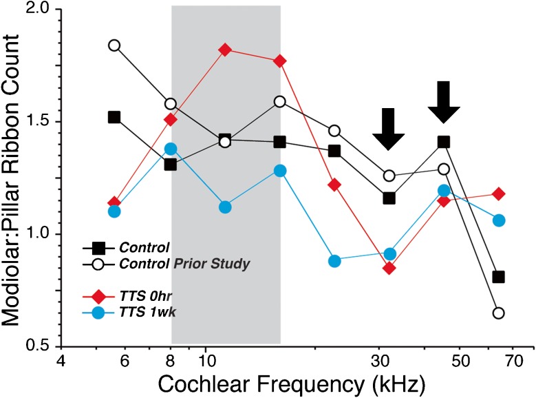
FIG. 8.
Neuropathic noise exposure decreases the number of synapses on the modiolar side of the IHC. Each graph is derived from the same dataset shown in Figure 3B by counting the number of synapses on the modiolar vs. the pillar sides of the IHC, as shown in Figures 4 and 7, and then expressing the difference as a ratio. Each point represents a single ensemble average across all the z-stacks at a given frequency region from all the ears in a given group. In control ears, there are always more synapses on the modiolar than the pillar side, except at the basal extreme of the cochlea (64 kHz). In the region of the noise-exposed ears, where the synaptic counts are maximally reduced, i.e., 32.0 and 45.2 kHz (arrows), there are slightly more synapses on the pillar than the modiolar side. To increase the robustness of the comparison, we include data from a separate set of control ears (group 2; n = 6) that was analyzed as part of a prior study of synaptic organization in mouse (Yin et al. 2014).

