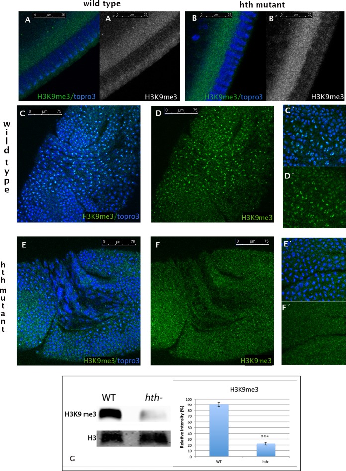
Fig 2. Tri-methylation of histone H3 in Lys9 is severely impaired in hth mutant embryos.
A, A´) Cycle 14 wild type embryo stained with a specific antibody that recognizes the H3K9me3 mark (green/grey). The signal is detected in the apical domain of the nuclei, which corresponds to the topro3 dense domain. B,B´) Cycle 14 hth mutant embryo stained as in A. Note the lack of signal in the nuclei. C, D) Stage 8 embryo stained as in A. High levels of H3K9me3 are observed in all the mitotic cells and lower levels are detected in the heterochromatin domains of the interphase nuclei (see inset C´, D´). E, F) In a stage 8 mutant embryo the levels of H3K9me3 are very much reduced when compared to wild type embryos (see inset F´). (Embryos were staged as in [61]). G) Western blot analysis of chromatin extracts confirms that the chromatin extracted from hth mutants show reduced levels of the H3K9me3 methyl mark. The graph represents the quantification of the signal observed. The western was performed three times with three different extractions (p-value: 0,00025212, calculated using T-Test). Error bars in the graph show the standard deviation and have being calculated as in Fig. 1 E. Anti-H3 was used as a loading control. Band quantification was done using ImageJ.

