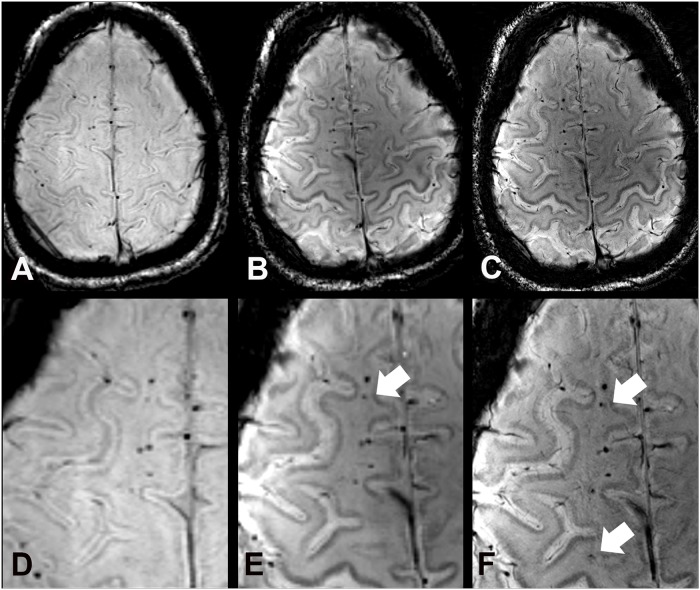
Fig 2. SWI images at 3 T (A), 7 T (B), and 7 T with high spatial resolution (C) of a 44 year old male study participant (case 5), who suffered from a traumatic brain injury in childhood.
Images show a line of traumatic microbleeds in the right frontal white matter. Compared to 3 T SW images (A, D) more hemorrhagic DAI lesions are depicted at 7 T with equal (B, E) and higher spatial resolution (C, F) (white arrows).

