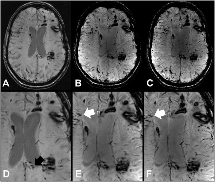
Fig 3. SWI images at 3 T (A, D) 7 T (B, E), and 7 T with high spatial resolution (C, F) of a 25 year old female DAI patient (case 7) survived a polytrauma with acute subdural hematoma, cerebral contusions, and DAI from a car accident with frontal collision.
In the splenium (black arrow) more TMB can be discriminated at 3 T SW images (A, D), which merge on 7 T SWI images due to the blooming effect. However, at 7 T SWI images (B, C, E, and F) demonstrate additional TMB in other brain regions like the right frontal white matter (white arrows).

